Category: Blog

Cusabio C-terminal 9xHis-tagged Recombinant

Abstract

We present the design of three expression vectors that can be used for rapid cloning of any blunt-ended DNA segment. Only a single set of oligonucleotides is required to perform target DNA amplification and cloning into all three vectors simultaneously. The DNA thus cloned can express a protein with or without a hexahistidine tag depending on the vector used. The expression occurs from the T7 promoter when transformed into E. coli BL21(DE3).

Two of the three plasmids have been designed to provide the expressed protein with 6 N- or C-terminal 9xHis-tagged Recombinant amino acids in tandem. The third plasmid, however, does not add any tag to the expressed protein. Cloning is rapidly achieved with the requirement of phosphorylation of the PCR product without any restriction digestion. Furthermore, clones generated can be confirmed with a one-step PCR reaction performed from bacterial colonies (generally referred to as “colony PCR”).

We show the cloning, expression and purification of Green Fluorescent Protein (GFP) as a proof of concept. Furthermore, we also show the cloning and expression of four sigma factors from Mycobacterium tuberculosis, further demonstrating the utility of the engineered plasmids. We strongly believe that the vectors and strategy we have developed will facilitate the rapid cloning and expression of any gene in E. coli BL21(DE3) with or without a hexahistidine tag.

Materials and methods

  • Bacterial strains, plasmids and growth conditions

Expression plasmids pET21b and pET15b were purchased from Novagen. EXPRESS chemically competent E. coli strains BL21(DE3) and XL1-blue were obtained from Lucigen and Stratagene respectively. E. coli bacterial strains were grown in Luria-Bertani medium (HiMedia), either as a liquid culture with constant shaking at 200 rpm or on a 1.5% agar plate at 37 °C. Cultures were always supplemented with 100 µg/ml ampicillin unless otherwise specified. All molecular biology methods and necessary precautionary measures were followed. Plasmid pSK01-NCHS was kindly provided by Soumya Kamilla, IISER Bhopal, India.

  • Reagents

Restriction endonucleases, Antarctic phosphatase, and T4 polynucleotide kinase (PNK) were obtained from New England Biolabs (NEB) and used according to the manufacturer’s instructions. T4 DNA ligase was obtained from Fermentas and used as suggested. Plasmid DNA purification and DNA gel extraction kits were purchased from Qiagen and Sigma, respectively. The 2 log DNA ladder was obtained from NEB for DNA electrophoresis on agarose gels. The PiNK Plus protein ladder was purchased from GeneDireX and used according to instructions in acrylamide gel electrophoresis experiments. All other reagents were purchased from Sigma.

  • GFP Expression and Zymography

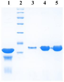
Plasmids carrying the GFP gene were transformed into E. coli BL21(DE3) cells. Each clone was grown at 37°C at 200 rpm in 5 ml of LB medium. At an optical density at 600nm (OD600), ~0.8, 0.5 ml of culture was removed and 1 mM IPTG (final concentration) was added to the remaining media for protein expression. Cultures were allowed to grow further for 3 hours. The bacteria in all these cases were collected by centrifugation and 50 µl of 8 M urea and 10 µl of 5X SDS gel loading buffer were added to each.

Cells were lysed by heating at 100°C for 5 min and 8 µl of each sample was subjected to 12% SDS-polyacrylamide gel electrophoresis (PAGE). After performing electrophoresis, the gel was incubated in 1% Triton X-100 for 2 hours and then photographed, to obtain a zymogram, with epi-illumination at 480 nm and SYBR Gold filter (485-655 nm) in a UVP. gel documentation system (UVP, LLC). Subsequently, the gel was fixed in a solution of 10% acetic acid, 1% trichloroacetic acid (TCA), and 40% methanol in water for 1 hour and then stained with Coomassie Brilliant Blue R-250 stain to visualize the protein bands.

GFP purification

Clones expressing GFP along with the His6 tag were grown at 37°C at 200 rpm in 250 ml of LB medium. The protein was expressed by the addition of 1 mM IPTG when the culture reached OD600 ~0.8. The induction was continued for 3 hours. Cells were then collected by centrifugation at 4 °C and resuspended in the lysis buffer (50 mM sodium phosphate buffer, 500 mM NaCl, 5 mM β-mercaptoethanol, 10 mM imidazole, 5% glycerol) supplemented with 10 µg/ml lysozyme (chicken egg white lysozyme; Sigma) and incubated on ice for 30 min.

Cells were then lysed by sonication and the lysate cleared by centrifugation. The supernatant was incubated with 250 µl bed volume of Ni-NTA (Nickel-Nitrilotriacetic acid) agarose (Qiagen), pre-equilibrated with the lysis buffer, for 1 hour with constant mixing at 4°C. The matrix was collected and washed with wash buffer (50mM sodium phosphate buffer, 1M NaCl, 5mM β-mercaptoethanol, 20mM imidazole, 5% glycerol). Protein elution was then carried out in 10 column volumes of elution buffer (50 mM sodium phosphate buffer, 500 mM NaCl, 5 mM β-mercaptoethanol, 200 mM imidazole, 5% glycerol) and fractions elution collected were analyzed at a rate of 12%. SDS-PAGE.

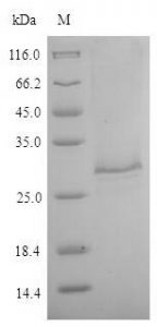
Cloning of Sigma Factors from Mycobacterium tuberculosis and their Expression Analysis

Four extracytoplasmic sigma factors (SigB, D, F and G) from M. tuberculosis were cloned into the designed PMS-QS vectors. Genes were amplified by PCR using the genomic DNA of M. tuberculosis H37Rv as a template (kindly provided by AstraZeneca, Bangalore) and the primers. The PCR products were extracted from the agarose gel and ligated into the vectors. linear PMS-QS. . The resulting products were transformed into E. coli XL1Blue and positive clones were identified by colony PCR. All clones were further confirmed by sequencing. Plasmids so constructed were transformed into E. coli BL21 (DE3) for protein expression as described above.

Results

In the present manuscript, we report the design and construction of “rapid series” PMS vectors. These vectors contain a restriction enzyme site that blunts the vector for cloning of the DNA fragments generated by PCR. The vector carries a T7 promoter and a ribosome binding site upstream of the cloning site with the appropriate addition of codons for six histidine amino acids.


Cusabio Cardiovascular Recombinants

Abstract

Cardiovascular Recombinant human erythropoietin (rHuEPO) treatment has solved the problem of anaemia in dialysis patients. However, its application to predialysis patients has raised some doubts about its effects on the progression of kidney disease and on the regulation of blood pressure (BP) and hemodynamics. We have prospectively studied for at least 6 months a group of 11 pre-dialysis patients treated with rHuEPO (initial dose, 1000 U subcutaneously three times a week). Clinical evaluation and biochemical and haematological measurements were performed once every 2 weeks.

Twenty-four-hour ambulatory BP monitoring, echocardiography, and determination of neurohumoral mediators of hemodynamics were performed once every 3 months. An adequate hematological response was found (hemoglobin, 11.7 +/- 0.4 g/dL v 9 +/- 0.3 g/dL) without changes in the progression of kidney disease. A decrease in cardiac output and an increase in total peripheral resistance were observed as anaemia improved. There was a trend toward decreased left ventricular (LV) thickness and a significant decrease in LV mass index (from 178.2 +/- 20.6 g/m2 to 147.3 +/- 20.6 g/m2). /m2).

Blood pressure control did not improve; In addition, in some patients, an increase in systolic values ​​was detected by ambulatory BP. Casual BP was apparently stable. Sequential determinations of neurohumoral mediators of hemodynamic substances (endothelin, renin, norepinephrine, epinephrine, dopamine) failed to explain these results. Ambulatory BP reveals worse control in some previously hypertensive patients and confirms the usefulness of this technique in the assessment of patients receiving erythropoietin treatment.

The trend toward regression of LV hypertrophy without better BP control confirms the role of anaemia among the multiple factors leading to LV hypertrophy in ESRD and opens therapeutic possibilities. Better BP control can potentially offset the beneficial effects that correcting anaemia would have on the cardiovascular system.

Purity: Greater than 90% as determined by SDS-PAGE.

Target Names: VEGFC

Uniprot No.: P49767

Research Area: Cancer

Alternative Names

Flt 4L; Flt4 ligand; FLT4 ligand DHM; Flt4-L; Flt4L; Vascular endothelial growth factor C; Vascular endothelial growth factor-related protein; Vascular endothelial growth factor-related protein; VEGF C; VEGF-C; Vegfc; VEGFC_HUMAN; PVR

Species: Homo sapiens (Human)

Source: E.coli

Expression Region: 112-227aa

Mol. Weight: 40.1kDa

Protein Length: Full Length of Mature Protein

Tag Info: N-terminal GST-tagged

Form: Liquid or Lyophilized powder

Note:

We will preferentially ship the format that we have in stock, however, if you have any special requirements for the format, please remark your requirement when placing the order, we will prepare according to your demand.

Buffer

If the delivery form is liquid, the default storage buffer is Tris/PBS-based buffer, 5%-50% glycerol.

Note: If you have any special requirements for the glycerol content, please remark when you place the order. If the delivery form is a lyophilized powder, the buffer before lyophilization is Tris/PBS-based buffer, 6% Trehalose, pH 8.0.

Reconstitution

We recommend that this vial be briefly centrifuged prior to opening to bring the contents to the bottom. Please reconstitute protein in deionized sterile water to a concentration of 0.1-1.0 mg/mL. We recommend adding 5-50% of glycerol (final concentration) and aliquot for long-term storage at -20°C/-80°C. Our default final concentration of glycerol is 50%. Customers could use it as a reference.

Cusabio Epigenetics & Nuclear Signaling Recombinants

Epigenetic modifications

Epigenetic changes can modify the activation of certain genes, but not the DNA sequence. Furthermore, chromatin proteins associated with DNA can be activated or silenced. This is the reason why the differentiated cells in a multicellular organism express only the genes that are necessary for their own activity. Histone modifications are epigenetic marks that affect gene expression patterns.

Abstract

Transcription, translation, and subsequent protein modification represent the transfer of genetic information from archival DNA to short-lived messenger RNA, usually with subsequent protein production. Although all cells in an organism contain essentially the same DNA, cell types and functions differ due to qualitative and quantitative differences in their gene expression.

Therefore, control of gene expression is at the core of differentiation and development. Epigenetics & Nuclear Signaling Recombinants processes, including DNA methylation, histone modification, and various RNA-mediated processes, are thought to influence gene expression primarily at the level of transcription; however, other steps in the process (eg translation) can also be epigenetically regulated. The following article will describe the role epigenetics is thought to play in influencing gene expression.

Epigenetics and gene expression

Transcription, translation, and subsequent protein modification represent the transfer of genetic information from archival DNA to short-lived messenger RNA, usually with subsequent protein production. Although all cells in an organism contain essentially the same DNA, cell types and functions differ due to qualitative and quantitative differences in their gene expression, and thus control of gene expression is at the core of differentiation. and the development.

Gene expression patterns that characterize differentiated cells are established during development and are maintained as cells divide by mitosis. Therefore, in addition to inheriting genetic information, cells inherit information that is not encoded in the DNA nucleotide sequence, and this has been called epigenetic information. Epigenetics has been defined as “the study of inherited mitotic (and potentially meiotic) alterations in gene expression that are not caused by changes in the DNA sequence” (Waterland, 2006).

However, some definitions of epigenetics are broader and do not necessarily encompass the heritability requirement. For example, the US National Institutes of Health (2009) in its recent initiative on epigenomics states that “epigenetics refers to both hereditary changes in gene activity and expression (in the progeny of cells or of individuals) as well long-term stable alterations”. in the transcriptional potential of a cell that is not necessarily heritable’.

Regardless of the exact definition, epigenetic processes that stably alter gene expression patterns (and/or mediate alterations in cell division) are believed to include: (1) cytosine methylation; (2) post-translational modification of histone proteins and chromatin remodelling; and (3) RNA-based mechanisms. Gene expression is a complex process involving numerous steps (Alberts et al., 2008) and although a detailed description of gene expression is beyond the scope of this review, we will briefly summarize the main steps to locate the subject of the review. , how epigenetics interacts with gene expression, in context.

The initial step in gene expression is the transcription of the DNA molecule into an exact copy of RNA. To initiate transcription, RNA polymerase binds to a particular region of DNA (the promoter) and begins to produce an mRNA strand that is complementary to one of the DNA strands. Post-transcriptional processing is critical: a methylated guanosine “cap” is added to the 5′ end of the transcribed RNA, while mRNA splicing occurs through a stepwise series of cleavage and ligation events that remove intron sequences and they join the exons in an appropriate way.

After splicing, the 3′ end of the mRNA is cleaved and a chain of adenosine residues, known as the polyA tail, is added in preparation for transport of the mRNA from the nucleus to the cytoplasm. At this stage, the mRNA is ready to bind to ribosomes for translation. In translation, polypeptides are synthesized sequentially stepwise from the N-terminus to the C-terminus through three distinct steps: initiation, elongation, and termination.

PaxView TB/NTM MPCR-ULFA Kit

Abstract

To determine whether patients with tuberculosis (TB) and nontuberculous mycobacteria (NTM) infection could be distinguished from each other with limited information, we compared patients with pulmonary TB and NTM during 2005-2006. Our finding that age, place of birth, and the presence of chronic obstructive pulmonary disease could differentiate TB and NTM disease could help in TB control efforts.

Keywords:

Bacteria, Mycobacterium tuberculosis, Mycobacterium avium, tuberculosis and other mycobacteria, pulmonary and respiratory infections, non-tuberculous mycobacteria, infection control, diagnosis, clearance.

General description

  • Non-tuberculous mycobacteria (NTM) are mycobacteria other than M. tuberculosis (the cause of tuberculosis) and M. leprae (the cause of leprosy). NTMs are also called atypical mycobacteria, mycobacteria other than tuberculosis (MOTT), or environmental mycobacteria.
  • Although anyone can get an NTM infection, NTMs are opportunistic pathogens that put some groups at higher risk, including those with underlying lung disease or suppressed immune systems. These pathogens are not normally spread from person to person. However, person-to-person transmission of M. abscessus has been reported in patients with cystic fibrosis.
  • NTMs are environmental organisms that can be found in soil, dust, and water, including natural water sources (such as lakes, rivers, and streams) and municipal water sources (such as the water that people drink or use). showering). NTM can form difficult-to-remove biofilms, which are collections of microorganisms that stick together and adhere to surfaces in moist environments, such as the inside of building plumbing.

Symptoms

1. NTMs can cause infections in a wide variety of body sites, most commonly the lungs and the following areas:

  • Skin and soft tissues (typically after surgery, trauma, injection of drugs or other substances)
  • Device-associated infections (eg, central line-associated bloodstream infections, exit-site infections, pacemaker pocket site infections, etc.)
  • Lymph nodes (more commonly in children)
  • Blood or other generally sterile places in the body (spread) (most commonly in immunocompromised patients, such as those with HIV or AIDS, but can also be due to invasive medical devices or procedures)

2. Symptoms can be vague and nonspecific, such as:

  • Fever
  • Weightloss
  • Night sweats
  • Decreased appetite
  • power loss

3. Other symptoms depend on the site of infection and may include cough, shortness of breath, blood in the sputum, and skin rashes.

Tuberculosis (TB) is a chronic bacterial disease that infects the lungs, kidneys, spine, and/or brain. TB is an airborne mycobacterium and can be spread from person to person, such as when an infected person coughs or sneezes. It can also cause an infection after a latent period in a person who was infected at an earlier time.

Without treatment, TB can cause serious complications in other parts of the body or even be fatal. Treatment requires months of adherence to various antibiotics, but most cases can be cured. Drug-resistant TB is much more difficult to cure and treatment takes much longer (9 to 18 months). Brigham and Women’s Hospital (BWH) works with the Massachusetts Department of Public Health, which subsidizes all diagnostic testing and treatment.

Surgery is used to diagnose and treat complications of TB. Surgical approaches to lung resection include thoracotomy and video-assisted thoracic surgery (VATS). For surgical treatment to be most effective, it must be preceded and followed by extensive antibacterial therapy.

Non-tuberculous mycobacterial (NTM) disease is a general term for a group of lung infections caused by exposure to mycobacteria found in soil and water. The mycobacteria that cause these diseases exclude those that cause tuberculosis and leprosy. NTM most commonly affects people with weakened immune systems due to other lung diseases, but healthy people with no history of lung disease can also be affected. If left untreated, the disease can become chronic and require ongoing treatment.

Despite medical and surgical advances, TB remains a global epidemic and its presentation requires an aggressive and coordinated approach. The Lung Center at BWH provides high-quality multidisciplinary care and research in the area of ​​TB and NTM treatment. Pulmonologists, thoracic surgeons, and infectious disease physicians who specialize in TB and NTM collaborate with other BWH specialists to provide patients with a highly informed diagnosis and consistent treatment plan.

What are the risk factors for TB and nontuberculous mycobacterial disease?

Tuberculosis and nontuberculous mycobacterial diseases affect people of all ages, genders, races, and income levels. The following groups face a higher risk:

  • People who live or work with other people who have TB, including hospital staff
  • People from countries where TB is prevalent
  • People who abuse alcohol or use intravenous drugs
  • people who smoke
  • Homeless people
  • Young children and the elderly
  • People without access to health care.
  • People in group settings, such as nursing homes.

Patients with weakened immune systems are particularly susceptible to both TB and nontuberculous mycobacterial disease. Some conditions that weaken the immune system and/or damage the lung structure include:

  • bronchiectasis
  • Chronic obstructive pulmonary disease (COPD) and emphysema
  • Cystic fibrosis
  • Human immunodeficiency virus (HIV)
  • Occupational and environmental lung diseases
  • lung transplant

Allergy Test for Animal (ANITIA Canine IgE)

Allergy tests and your pet

Just like us, pets can also experience allergies. Spring pollen, irritating shampoos, and even food allergies can be a problem for pets. Fortunately, allergy tests can often identify the cause of your pet’s reaction.

How is it done?

Your pet can be screened for a list of environmental allergens with a simple blood test. The sample is sent to a laboratory where tests will determine the cause and severity of your pet’s allergy.

How does it help?

The best treatment for an allergy is to avoid contact with the offending substance. However, this is not always possible. In many cases, intermittent treatment is necessary. Steroids, antibiotics, antihistamines, special shampoos, or supplements may be used to treat allergies. However, if used for too long, steroids can have adverse side effects.

Antibiotics and antihistamines can be a considerable expense. Allergy-positive pets may benefit from desensitizing vaccines specially formulated for your pet. The goal is to control your pet’s allergy and minimize the need for additional medication.

How successful is it?

Dogs and cats, like people, are individuals. No two dogs or cats are exactly alike and neither are their allergies. Therefore, some patients will respond better than others. Allergy shots are not a quick cure for your pet’s allergies. Rather, they are safer and, in many cases, cheaper than alternative treatment methods.

What is the treatment process?

The initial blood test is taken at a normal veterinary appointment. When the treatment vials are tested by a laboratory, an injection regimen is started. If your pet develops clinical symptoms of his allergy during treatment, they can be controlled through the use of antibiotics or shampoos. The use of steroids during desensitization is avoided. Your Greencross veterinarian will administer the injections during scheduled, one-on-one visits. Talk to your local Greencross veterinarians if you are concerned about allergies in your pet.

Allergen:

Something to which the immune system develops an allergy; they are usually proteins in things like pollen, dust mites, and some moulds. Antibody: A specialized protein called immunoglobulin (Ig). There are many subtypes, including those that are responsible for allergic reactions, IgE. Gold standard: For the purpose of our discussion: a diagnostic test that is considered to be the most reliable.

When evaluating the usefulness or reliability of a new or different test, that test is compared to the “gold standard” test. Hyposensitization Injections – Another name for allergy shots or allergy shots. These injections reduce the patient’s sensitivity to allergens. IgE: The allergic antibody; type E immunoglobulin.

Allergy testing is done to determine which allergens may be responsible for a pet’s allergy symptoms. Allergy tests do this by looking for the presence of specific allergy-type antibodies (IgE) to common allergens. Two areas of the body are commonly tested for the presence of specific IgE: the skin and also the blood.

The intradermal allergy test (or allergy skin test) is considered the gold standard of allergy testing. It is very similar to the allergy skin test that is done on humans. In pets, it consists of injecting small amounts of allergens in an organized way into the skin. When a pet produces IgE that is specific to the injected allergen during testing, the IgE produced by the pet causes the skin to redden and swell exactly where the allergen was injected.

A scoring system is used to differentiate positive from negative injection sites. Training and experience are needed to test injections correctly. Skin allergy tests, like blood allergy tests, are reliable only for non-food allergens such as pollen, dust mites, and mould.

Does your dog or cat scratch and groom itself too much?

Dog and cat allergy symptoms are distressing for your pet. Skin allergies are one of the most common skin conditions we see in dogs and cats and can have a negative impact on your pet’s life. Atopic dermatitis is a reaction to environmental aeroallergens such as pollen from grasses, weeds, trees, mould spores, and house dust mites.

What are the common symptoms of cat allergies?

  • Belly and sides too trimmed
  • excessive hair loss
  • Itchy crusty rash
  • Itchy, moist, red raised lesions in the groin or armpit region
  • Severe scratching of the head, ears, and neck
  • Greasy and dishevelled coat
  • Changing diet doesn’t make any difference.

What are common dog allergy symptoms?

  • Itching, most commonly around the nose, eyes, ear flaps, armpits, groin, and legs (especially if the itching affects the feet, face, ears, buttocks, legs, or areas that come in contact with the ground when the dog lies down)
  • Recurring skin or ear infections
  • Inflamed skin prone to secondary infections with bacteria and yeast (as a result of chewing, licking, rubbing, and scratching)
  • If your dog becomes “smelly” within a few days of being washed
  • If symptoms started or get worse in hot weather
  • If your dog gets itchy after touching grass
  • If your dog is itchy after being washed
  • If your dog has persistently oily skin
  • If a diet change makes no difference

How do we diagnose atopic skin disease?

We identify allergies by intradermal allergy testing. This involves cutting a strand of hair from one side and pricking the skin with a small amount of purified allergen extract. If the skin swells thirty minutes after the injection, it means that an allergy has been detected. Another way to identify allergens is to perform blood serology tests. Using the results of intradermal and blood tests provides a more accurate allergy profile for your pet and a better outcome with immunotherapy.

*Please note: Intradermal allergy testing is affected by a number of medications, including antihistamines and corticosteroids (cortisone tablets, injections, lotions, eye and ear drops). It is important that your pet is taken off treatment prior to testing. Talk to our technicians about medication withdrawal times for your dog and cat.

How do we treat atopic dermatitis?

The best treatment for allergies is usually immunotherapy (desensitization) or avoiding allergy triggers. Immunotherapy involves creating a vaccine for your pet. Once we have identified what your pet is allergic to, we formulate an allergy vaccine (immunotherapy) that contains the allergens your pet has reacted to. As the vaccine starts to work, there will be fewer allergic signs like infections and itching. We prefer to use immunotherapy over medication, as it minimizes subsequent reactions.

COVID-19 N-Antigen

Objectives:

COVID-19 emerged and rapidly spread throughout the world. Testing strategies focussing on patients with COVID-19 require assays that are high-throughput, low-risk of infection, and with small sample volumes. Antigen surveillance can be used to identify exposure to pathogens and measure acute infections.

Methods:

A total of 914 serum samples, collected from 309 currently infected COVID-19 patients, 48 recovered ones, and 410 non-COVID-19 patients, were used to measure N protein antigen levels by a chemiluminescent immunoassay. Diagnostic performances were analyzed in different periods after onset.

  • Patients, samples and data collection

Consecutive patients with COVID-19 who were presented or admitted to Zhongnan Hospital, Wuhan University from January 28 to March 10, 2020, were included to test for SARS-COV-2 N-protein antigen and specific antibodies in serum. COVID-19 and recovered patients enrolled in this study were diagnosed according to the diagnostic guideline published by the National Health Commission.

Healthy volunteers and other virus-infected patients who were examined or admitted to Zhongnan Hospital, Wuhan University, from November 18, 2020, to December 6, 2020, were enrolled in the control group. Exclusion criteria for controls were as follows: (a) SARS-COV-2 RNA positive in throat swab, (b) patients with poor basic clinical data. All throat swabs and venous blood samples were collected and processed at Zhongnan Hospital of Wuhan University.

The remaining sera were collected and stored at -80°C for SARS-CoV-2 specific antibody and N-protein antigen testing. Clinical characteristics, laboratory findings, and results were collected from the medical record. electronics. The study was reviewed and approved by the Ethics Committee of Zhongnan Hospital, Wuhan University. All study targets have signed informed consent.

  • Real-time RT-PCR assay for SARS-CoV-2 RNA

Throat swabs were collected from COVID-19 patients for SARS-CoV-2 RNA testing. First, total RNAs were extracted from the swab within 3 h using a respiratory sample RNA isolation kit (Zhongzhi, Wuhan, China). Briefly, 40 μL of cell lysis solution was transferred to a collection tube consisting of a swab followed by vortexing for 30 s. After incubation at room temperature for 15 min, the collection tube was centrifuged at 1000 rpm/min for 5 min.

The suspension was used as a template for amplification using real-time reverse transcriptase-polymerase chain reaction (RT-PCR) assay kits (Daan Gene, Guangzhou, China). Two target genes, SARS-CoV-2 nucleocapsid protein (N) and open reading frame 1ab (ORF1ab), were simultaneously amplified and detected during real-time RT-PCR assay. The real-time RT-PCR reaction system volume was 25 μL, including 2 μL of template, 3 μL of pure water, 17 μL of mix A, and 3 μL of mix B.

Each amplification was performed in an Eppendorf tube. by ABI prism 7500 (Thermo Fisher Scientific, Waltham, MA, USA). The reaction conditions were as follows: transcription at 50°C for 15 min and pre-denaturation at 95°C for 15 min, followed by 45 cycles of denaturation at 94°C for 15 s and extension at 55°C for 45 s. Fluorescence was collected at regular intervals during each extension phase. The lower detection limit of the real-time RT-PCR assay for two target genes was 500 copies/mL. According to the manufacturer’s recommendation, a cycle threshold value (Ct) of <40 was defined as positive.

  • Chemiluminescence immunoassay to analyze SARS-CoV-2 N serum antigen

Serum SARS-CoV-2 protein N antigen was determined by double-antibody sandwich chemiluminescence immunoassay using an iFlash immunoassay analyzer (Shenzhen Yhlo Biotech Co., Ltd, Shenzhen, China). Paramagnetic carboxylated microparticles (Thermo Scientific) were coated with one of 10 candidate-specific antibodies (Shenzhen YHLO Biotech Co., Ltd, Shenzhen, China) by cross-linking N-ethyl-N’-(3-dimethyl aminopropyl)carbodiimide (Thermo Scientific) for protein N antigen-capture as described above. Another antibody was conjugated with NSP-DMAE-NHS (Maxchemtech) for antigen detection.

SARS-CoV-2 recombination nucleocapsid protein (Shenzhen YHLO Biotech) dissolved in healthy human serum was used as a calibrator. Tests can be run after calibration. In the test, paramagnetic carboxylated microparticles coated with the capture antibody bound N-protein antigens. After washing away unbound material, the antibody N-protein antigen-capture antibody compounds reacted with the acridinium-labelled antibody.

The mixture was kept in a tube under a magnetic field. And then the preactivation and activation solution were added to calculate the N-protein antigen based on the resulting relative light units (RLU) through a 2-point calibration curve.

  • Statistic analysis

Statistical analyzes were performed with IBM SPSS version 23.0 software. Normal distribution continuous data were presented as mean ± standard deviation (SD) and skewed distribution continuous data were presented as median and range. The student’s t-test or non-parametric test was used for the comparison of continuous data. The chi-square test was used to analyze categorical data. Diagnostic sensitivity and specificity were calculated using the receiver operating characteristic (ROC) curve and the area under the curve (AUC). p ≤ 0.05 was considered statistically significant.

Results:

There was a high level of N protein antigen in COVID-19 patients (0.56 COI), compared to the recovered patients (0.12 COI) and controls (0.19 COI). In receiver-operating characteristic curve analysis, the area under the curve of serum N protein antigen was 0.911 in the first week after onset. In this period, the Sensitivity and specificity of serologic N protein antigen testing was 76.27 and 98.78%. Diagnosis performance of specific antibodies became better from the third week after onset. Subgroup analysis suggested that severe patients had higher levels of antigens than mild patients.

Conclusions:

A high level of serum antigen suggested early infection and serious illness. Serum N protein antigen testing by chemiluminescence immunoassay is considered a viable assay used to improve diagnostic sensitivity for current patients.

Keywords

Antigen, antigenemia, Blood, COVID-19, Diagnosis, Plasma, SARS-CoV-2, Serum

HiiScript ii U+ One Step qRT-PCR Probe Kit

Product introduction

The dUTP/UDG anti-contamination system is a very effective means of controlling the contamination of PCR amplification products. In PCR detection reagents using DNA as a template, it has gradually become a mandatory standard.

However, the commonly used E. coli UDG still has high activity at the usual reverse transcription temperature (42-55℃), which will degrade the cDNA, thus reducing the sensitivity of One Step RT-PCR/qPCR. The Hiscript II U+ One-Step qRT-PCR Probe Kit contains an optimized ratio of dUTP/dNTP mix and heat-labile UDG derived from psychrophilic marine bacteria.

Heat-labile UDG has high activity at room temperature and can completely degrade U-containing double-stranded DNA during the mixing process of the reaction system. When the reaction system is heated to 50-55°C (the optimal HiScript II reaction temperature), the heat-labile UDG is rapidly and completely inactivated, maintaining cDNA integrity and ensuring that detection sensitivity is not compromised. be affected.

Storage conditions

Store at -20℃

Product Advantages

  • Support multiple probe detection;
  • UDG anti-contamination system: Heat-labile UDG rapidly degrades U-containing double-stranded DNA contaminants at room temperature. Heat-labile UDG is rapidly and completely inactivated at 50-55°C to maintain cDNA integrity;
  • Ultra-high detection sensitivity: single-digit template copies or 0.1 pg of total RNA can be detected
  • Ultra-high amplification specificity: AceTaq® DNA polymerase based on a hot chemical start, enzyme activity completely blocked before 95°C, and equipped with a patented specific promoting factor Exactor, which makes the amplification more specific;

ZENA SARS-CoV-2 Direct Detection Kit

Abstract

An epidemic of respiratory disease caused by severe acute respiratory syndrome coronavirus 2 (SARS-CoV-2) began in China and has spread to other countries. Real-time reverse transcriptase-polymerase chain reaction (RRT-PCR ) from nasopharyngeal swabs has generally been used to confirm the clinical diagnosis. However, it is unknown whether the virus can be detected in samples from other sites and thus may be transmitted in ways other than respiratory droplets.

Methods

We investigated the biodistribution of SARS-CoV-2 among different tissues of hospitalized patients with coronavirus disease 2019 (COVID-19) diagnosed based on symptoms and radiology and confirmed by SARS-CoV-2 detection. This study was approved by the ethics committees of the participating hospitals, waiving informed consent.

Patients were included with samples collected according to clinical indications from 3 hospitals in Hubei and Shandong provinces and Beijing, China, from January 1 to February 17, 2020. Pharyngeal swabs were collected from most patients at 1 to 3 days after hospital admission.

Blood, sputum, stool, urine, and nasal samples were collected throughout the illness. Bronchoalveolar lavage fluid and fiberoptic brush biopsy samples were taken from patients with severe disease or undergoing mechanical ventilation. RNA was extracted from clinical samples and determined by RRT-PCR targeting the SARS-CoV-2 open reading frame gene 1ab as described previously.2 RRT-PCR cycle threshold values ​​were used as indicators of the SARS-CoV copy number.

RNA in samples with lower cycle threshold values ​​corresponding to higher viral copy numbers. A cycle threshold value less than 40 is interpreted as positive for SARS-CoV-2 RNA. Four high copy number SARS-CoV-2 positive faecal samples were cultured and then electron microscopy was performed to detect the live virus. Patterns were explored in a subgroup of patients with multiple samples collected during hospitalization.

Results

A total of 1,070 samples were collected from 205 COVID-19 patients who had a mean age of 44 years (range, 5-67 years) and 68% were men. Most of the patients presented fever, dry cough and fatigue; 19% of patients had severe disease. Bronchoalveolar lavage fluid samples showed the highest positive rates (14 of 15; 93%), followed by sputum (72 of 104; 72%), nasal swabs (5 of 8; 63%), fiberoptic brush biopsy (6 of 13; 46%), throat swabs (126 of 398; 32%), stool (44 of 153; 29%), and blood (3 of 307; 1%). None of the 72 urine samples was positive.

Mean cycle threshold values ​​for all sample types were greater than 30 (<2.6 × 10⁴ copies/mL), except for nasal swabs with a mean cycle threshold value of 24.3 (1.4 × 106 copies/mL), indicating higher viral loads.

Twenty patients had 2 to 6 specimens collected simultaneously. Viral RNA was detected in individual samples from 6 patients (respiratory samples, faeces or blood), while 7 patients excreted virus in respiratory tract samples and faeces (n = 5) or blood (n = 2). Live SARS-CoV-2 was observed in the stool sample of 2 patients who did not have diarrhoea.

Discussion

In this study, SARS-CoV-2 was detected in multi-site samples from 205 COVID-19 patients, with lower respiratory tract samples testing positive for the virus more frequently. Importantly, the live virus was detected in faeces, implying that SARS-CoV-2 can be transmitted via the faeces. A small percentage of blood samples had positive results on the PCR test, suggesting that the infection can sometimes be systemic.

Respiratory and extra-respiratory transmission of the virus may help explain the rapid spread of the disease. In addition, multi-site sample testing can improve sensitivity and reduce false-negative results. Two smaller studies reported the presence of SARS-CoV-2 in anal or oral swabs and blood from 16 patients in Hubei province, and the viral load in throat swabs and sputum from 17 confirmed cases.

Limitations of this study include that some patients did not have detailed clinical information available, so the data could not be correlated with symptoms or disease course, and that the number of some sample types was small. Further investigation of patients with detailed temporal and symptom data and consecutively collected samples from different sites is warranted.

Powered by WordPress & Theme by Anders Norén