Day: February 1, 2022

Cusabio C-terminal 9xHis-tagged Recombinant

Abstract

We present the design of three expression vectors that can be used for rapid cloning of any blunt-ended DNA segment. Only a single set of oligonucleotides is required to perform target DNA amplification and cloning into all three vectors simultaneously. The DNA thus cloned can express a protein with or without a hexahistidine tag depending on the vector used. The expression occurs from the T7 promoter when transformed into E. coli BL21(DE3).

Two of the three plasmids have been designed to provide the expressed protein with 6 N- or C-terminal 9xHis-tagged Recombinant amino acids in tandem. The third plasmid, however, does not add any tag to the expressed protein. Cloning is rapidly achieved with the requirement of phosphorylation of the PCR product without any restriction digestion. Furthermore, clones generated can be confirmed with a one-step PCR reaction performed from bacterial colonies (generally referred to as “colony PCR”).

We show the cloning, expression and purification of Green Fluorescent Protein (GFP) as a proof of concept. Furthermore, we also show the cloning and expression of four sigma factors from Mycobacterium tuberculosis, further demonstrating the utility of the engineered plasmids. We strongly believe that the vectors and strategy we have developed will facilitate the rapid cloning and expression of any gene in E. coli BL21(DE3) with or without a hexahistidine tag.
Materials and methods

  • Bacterial strains, plasmids and growth conditions

Expression plasmids pET21b and pET15b were purchased from Novagen. EXPRESS chemically competent E. coli strains BL21(DE3) and XL1-blue were obtained from Lucigen and Stratagene respectively. E. coli bacterial strains were grown in Luria-Bertani medium (HiMedia), either as a liquid culture with constant shaking at 200 rpm or on a 1.5% agar plate at 37 °C. Cultures were always supplemented with 100 µg/ml ampicillin unless otherwise specified. All molecular biology methods and necessary precautionary measures were followed. Plasmid pSK01-NCHS was kindly provided by Soumya Kamilla, IISER Bhopal, India.

  • Reagents

Restriction endonucleases, Antarctic phosphatase, and T4 polynucleotide kinase (PNK) were obtained from New England Biolabs (NEB) and used according to the manufacturer’s instructions. T4 DNA ligase was obtained from Fermentas and used as suggested. Plasmid DNA purification and DNA gel extraction kits were purchased from Qiagen and Sigma, respectively. The 2 log DNA ladder was obtained from NEB for DNA electrophoresis on agarose gels. The PiNK Plus protein ladder was purchased from GeneDireX and used according to instructions in acrylamide gel electrophoresis experiments. All other reagents were purchased from Sigma.

  • GFP Expression and Zymography

Plasmids carrying the GFP gene were transformed into E. coli BL21(DE3) cells. Each clone was grown at 37°C at 200 rpm in 5 ml of LB medium. At an optical density at 600nm (OD600), ~0.8, 0.5 ml of culture was removed and 1 mM IPTG (final concentration) was added to the remaining media for protein expression. Cultures were allowed to grow further for 3 hours. The bacteria in all these cases were collected by centrifugation and 50 µl of 8 M urea and 10 µl of 5X SDS gel loading buffer were added to each.

Cells were lysed by heating at 100°C for 5 min and 8 µl of each sample was subjected to 12% SDS-polyacrylamide gel electrophoresis (PAGE). After performing electrophoresis, the gel was incubated in 1% Triton X-100 for 2 hours and then photographed, to obtain a zymogram, with epi-illumination at 480 nm and SYBR Gold filter (485-655 nm) in a UVP. gel documentation system (UVP, LLC). Subsequently, the gel was fixed in a solution of 10% acetic acid, 1% trichloroacetic acid (TCA), and 40% methanol in water for 1 hour and then stained with Coomassie Brilliant Blue R-250 stain to visualize the protein bands.
GFP purification

Clones expressing GFP along with the His6 tag were grown at 37°C at 200 rpm in 250 ml of LB medium. The protein was expressed by the addition of 1 mM IPTG when the culture reached OD600 ~0.8. The induction was continued for 3 hours. Cells were then collected by centrifugation at 4 °C and resuspended in the lysis buffer (50 mM sodium phosphate buffer, 500 mM NaCl, 5 mM β-mercaptoethanol, 10 mM imidazole, 5% glycerol) supplemented with 10 µg/ml lysozyme (chicken egg white lysozyme; Sigma) and incubated on ice for 30 min.

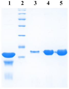
Cells were then lysed by sonication and the lysate cleared by centrifugation. The supernatant was incubated with 250 µl bed volume of Ni-NTA (Nickel-Nitrilotriacetic acid) agarose (Qiagen), pre-equilibrated with the lysis buffer, for 1 hour with constant mixing at 4°C. The matrix was collected and washed with wash buffer (50mM sodium phosphate buffer, 1M NaCl, 5mM β-mercaptoethanol, 20mM imidazole, 5% glycerol). Protein elution was then carried out in 10 column volumes of elution buffer (50 mM sodium phosphate buffer, 500 mM NaCl, 5 mM β-mercaptoethanol, 200 mM imidazole, 5% glycerol) and fractions elution collected were analyzed at a rate of 12%. SDS-PAGE.

Cloning of Sigma Factors from Mycobacterium tuberculosis and their Expression Analysis

Four extracytoplasmic sigma factors (SigB, D, F and G) from M. tuberculosis were cloned into the designed PMS-QS vectors. Genes were amplified by PCR using the genomic DNA of M. tuberculosis H37Rv as a template (kindly provided by AstraZeneca, Bangalore) and the primers. The PCR products were extracted from the agarose gel and ligated into the vectors. linear PMS-QS. . The resulting products were transformed into E. coli XL1Blue and positive clones were identified by colony PCR. All clones were further confirmed by sequencing. Plasmids so constructed were transformed into E. coli BL21 (DE3) for protein expression as described above.

Results

In the present manuscript, we report the design and construction of “rapid series” PMS vectors. These vectors contain a restriction enzyme site that blunts the vector for cloning of the DNA fragments generated by PCR. The vector carries a T7 promoter and a ribosome binding site upstream of the cloning site with the appropriate addition of codons for six histidine amino acids.


 

Cusabio Cardiovascular Recombinants

Abstract

Cardiovascular Recombinant human erythropoietin (rHuEPO) treatment has solved the problem of anaemia in dialysis patients. However, its application to predialysis patients has raised some doubts about its effects on the progression of kidney disease and on the regulation of blood pressure (BP) and hemodynamics. We have prospectively studied for at least 6 months a group of 11 pre-dialysis patients treated with rHuEPO (initial dose, 1000 U subcutaneously three times a week). Clinical evaluation and biochemical and haematological measurements were performed once every 2 weeks.

Twenty-four-hour ambulatory BP monitoring, echocardiography, and determination of neurohumoral mediators of hemodynamics were performed once every 3 months. An adequate hematological response was found (hemoglobin, 11.7 +/- 0.4 g/dL v 9 +/- 0.3 g/dL) without changes in the progression of kidney disease. A decrease in cardiac output and an increase in total peripheral resistance were observed as anaemia improved. There was a trend toward decreased left ventricular (LV) thickness and a significant decrease in LV mass index (from 178.2 +/- 20.6 g/m2 to 147.3 +/- 20.6 g/m2). /m2).

Blood pressure control did not improve; In addition, in some patients, an increase in systolic values ​​was detected by ambulatory BP. Casual BP was apparently stable. Sequential determinations of neurohumoral mediators of hemodynamic substances (endothelin, renin, norepinephrine, epinephrine, dopamine) failed to explain these results. Ambulatory BP reveals worse control in some previously hypertensive patients and confirms the usefulness of this technique in the assessment of patients receiving erythropoietin treatment.

The trend toward regression of LV hypertrophy without better BP control confirms the role of anaemia among the multiple factors leading to LV hypertrophy in ESRD and opens therapeutic possibilities. Better BP control can potentially offset the beneficial effects that correcting anaemia would have on the cardiovascular system.

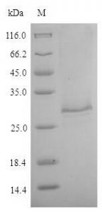
Purity: Greater than 90% as determined by SDS-PAGE.

Target Names: VEGFC

Uniprot No.: P49767

Research Area: Cancer

Alternative Names

Flt 4L; Flt4 ligand; FLT4 ligand DHM; Flt4-L; Flt4L; Vascular endothelial growth factor C; Vascular endothelial growth factor-related protein; Vascular endothelial growth factor-related protein; VEGF C; VEGF-C; Vegfc; VEGFC_HUMAN; PVR

Species: Homo sapiens (Human)

Source: E.coli

Expression Region: 112-227aa

Mol. Weight: 40.1kDa

Protein Length: Full Length of Mature Protein

Tag Info: N-terminal GST-tagged

Form: Liquid or Lyophilized powder

Note:

We will preferentially ship the format that we have in stock, however, if you have any special requirements for the format, please remark your requirement when placing the order, we will prepare according to your demand.

Buffer

If the delivery form is liquid, the default storage buffer is Tris/PBS-based buffer, 5%-50% glycerol.

Note: If you have any special requirements for the glycerol content, please remark when you place the order. If the delivery form is a lyophilized powder, the buffer before lyophilization is Tris/PBS-based buffer, 6% Trehalose, pH 8.0.

Reconstitution

We recommend that this vial be briefly centrifuged prior to opening to bring the contents to the bottom. Please reconstitute protein in deionized sterile water to a concentration of 0.1-1.0 mg/mL. We recommend adding 5-50% of glycerol (final concentration) and aliquot for long-term storage at -20°C/-80°C. Our default final concentration of glycerol is 50%. Customers could use it as a reference.

Cusabio Epigenetics & Nuclear Signaling Recombinants

Epigenetic modifications

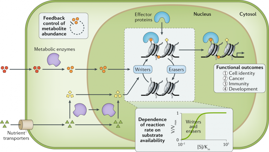
Epigenetic changes can modify the activation of certain genes, but not the DNA sequence. Furthermore, chromatin proteins associated with DNA can be activated or silenced. This is the reason why the differentiated cells in a multicellular organism express only the genes that are necessary for their own activity. Histone modifications are epigenetic marks that affect gene expression patterns.

Abstract

Transcription, translation, and subsequent protein modification represent the transfer of genetic information from archival DNA to short-lived messenger RNA, usually with subsequent protein production. Although all cells in an organism contain essentially the same DNA, cell types and functions differ due to qualitative and quantitative differences in their gene expression.

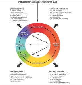
Therefore, control of gene expression is at the core of differentiation and development. Epigenetics & Nuclear Signaling Recombinants processes, including DNA methylation, histone modification, and various RNA-mediated processes, are thought to influence gene expression primarily at the level of transcription; however, other steps in the process (eg translation) can also be epigenetically regulated. The following article will describe the role epigenetics is thought to play in influencing gene expression.

Epigenetics and gene expression

Transcription, translation, and subsequent protein modification represent the transfer of genetic information from archival DNA to short-lived messenger RNA, usually with subsequent protein production. Although all cells in an organism contain essentially the same DNA, cell types and functions differ due to qualitative and quantitative differences in their gene expression, and thus control of gene expression is at the core of differentiation. and the development.

Gene expression patterns that characterize differentiated cells are established during development and are maintained as cells divide by mitosis. Therefore, in addition to inheriting genetic information, cells inherit information that is not encoded in the DNA nucleotide sequence, and this has been called epigenetic information. Epigenetics has been defined as “the study of inherited mitotic (and potentially meiotic) alterations in gene expression that are not caused by changes in the DNA sequence” (Waterland, 2006).

However, some definitions of epigenetics are broader and do not necessarily encompass the heritability requirement. For example, the US National Institutes of Health (2009) in its recent initiative on epigenomics states that “epigenetics refers to both hereditary changes in gene activity and expression (in the progeny of cells or of individuals) as well long-term stable alterations”. in the transcriptional potential of a cell that is not necessarily heritable’.

Regardless of the exact definition, epigenetic processes that stably alter gene expression patterns (and/or mediate alterations in cell division) are believed to include: (1) cytosine methylation; (2) post-translational modification of histone proteins and chromatin remodelling; and (3) RNA-based mechanisms. Gene expression is a complex process involving numerous steps (Alberts et al., 2008) and although a detailed description of gene expression is beyond the scope of this review, we will briefly summarize the main steps to locate the subject of the review. , how epigenetics interacts with gene expression, in context.

The initial step in gene expression is the transcription of the DNA molecule into an exact copy of RNA. To initiate transcription, RNA polymerase binds to a particular region of DNA (the promoter) and begins to produce an mRNA strand that is complementary to one of the DNA strands. Post-transcriptional processing is critical: a methylated guanosine “cap” is added to the 5′ end of the transcribed RNA, while mRNA splicing occurs through a stepwise series of cleavage and ligation events that remove intron sequences and they join the exons in an appropriate way.

After splicing, the 3′ end of the mRNA is cleaved and a chain of adenosine residues, known as the polyA tail, is added in preparation for transport of the mRNA from the nucleus to the cytoplasm. At this stage, the mRNA is ready to bind to ribosomes for translation. In translation, polypeptides are synthesized sequentially stepwise from the N-terminus to the C-terminus through three distinct steps: initiation, elongation, and termination.

Powered by WordPress & Theme by Anders Norén